Highlights
- Emerging trends in aortic valve disease management will shape clinical practice by 2025.
- Advances in diagnostic tools enable personalized treatment approaches for improved patient outcomes.
Summary
Understanding Aortic Valve Conditions: Key Insights for 2025 provides a comprehensive overview of the anatomy, epidemiology, pathophysiology, diagnosis, and management of aortic valve diseases, with an emphasis on recent advances and emerging trends shaping clinical practice by 2025. The aortic valve, a critical cardiac structure regulating blood flow from the left ventricle to the aorta, is subject to various pathological conditions, primarily aortic stenosis and aortic regurgitation, which can significantly impair cardiovascular function and patient outcomes.
Aortic valve diseases display considerable global variation in prevalence and etiology. In high-income countries, degenerative calcific aortic stenosis predominates, especially among the elderly, whereas rheumatic heart disease remains a major cause in low-income regions, often affecting younger populations. The bicuspid aortic valve, a common congenital anomaly, contributes to early-onset valvular dysfunction and presents unique clinical challenges. These demographic and geographic differences highlight the importance of tailored diagnostic and therapeutic approaches informed by evolving epidemiological data.
Diagnostic evaluation integrates clinical assessment with multimodality imaging techniques, including transthoracic echocardiography, cardiac magnetic resonance imaging, and computed tomography, supported by invasive hemodynamic measurements when indicated. Advances in biomarker research and imaging technologies have enhanced disease characterization and risk stratification, facilitating more personalized care pathways.
Treatment strategies have significantly evolved, with surgical aortic valve replacement (SAVR) remaining the standard for many patients, while transcatheter aortic valve replacement (TAVR) has expanded its role across diverse risk profiles and valve pathologies. Despite progress, challenges persist, particularly in managing aortic regurgitation via transcatheter methods due to anatomical complexities. Current clinical guidelines emphasize multidisciplinary decision-making that balances patient-specific factors such as comorbidities, frailty, and life expectancy to optimize timing and modality of intervention. Ongoing research focuses on improving device technologies, refining early intervention criteria, and exploring pharmacological therapies to address unmet needs in aortic valve disease management.
Anatomy and Physiology of the Aortic Valve
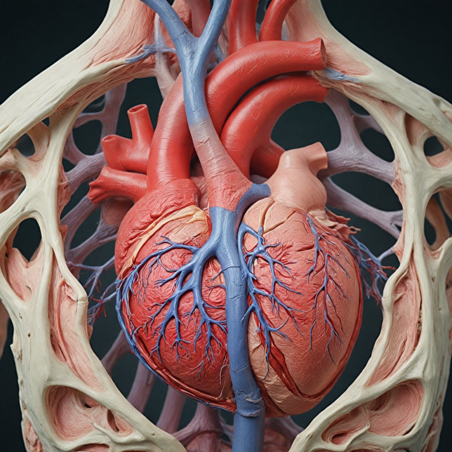
The aortic valve is a vital structure within the heart, situated between the left ventricle and the aorta, playing a critical role in the circulation of oxygenated blood throughout the body. It is one of the four heart valves and is often referred to as the aortic semilunar valve due to its distinctive semilunar shape.
Structure
The valve typically comprises three leaflets, also known as cusps, which are made predominantly of collagen. These leaflets are named according to the coronary arteries that arise from the corresponding sinuses of Valsalva: the left coronary cusp, right coronary cusp, and non-coronary cusp. However, variations in anatomy can lead to discrepancies in naming conventions, with some sources alternatively designating them as left, right, and posterior cusps or left posterior, anterior, and right posterior cusps. Each leaflet is connected to the aortic root through the aortic annulus and suspended by fibrous structures called commissures. The sinuses, which are wider than both the left ventricular outflow tract and the ascending aorta, create spaces behind each cusp that accommodate the origin of coronary arteries in two of the sinuses.
Supporting Fibrous Structures
Between the bases of the aortic valve sinuses lie the interleaflet fibrous triangles, also known as interleaflet triangles or intervalvular trigones. These fibrous triangles are anchored to the left ventricular wall and act as a boundary between the extracardiac space and the left ventricular cavity, extending up to the sinotubular junction. One particular interleaflet fibrous triangle, located between the right and left coronary sinuses, attaches to the septal portion of the right ventricular outflow tract and faces the pulmonary valve.
Function
The aortic valve ensures unidirectional blood flow from the left ventricle into the aorta, preventing backflow during ventricular diastole. During systole, the leaflets open widely to allow oxygen-rich blood to exit the heart and enter the systemic circulation. They then close tightly during diastole to prevent regurgitation back into the left ventricle, maintaining efficient cardiac function.
Epidemiology and Demographics
Aortic valve disease exhibits significant variability in epidemiology and demographics across different regions and populations. In developed nations, the condition predominantly affects the elderly, often presenting alongside other comorbidities such as chronic hypertension and atherosclerosis. Conversely, in low-income countries, valvular disease is more commonly seen in younger individuals, largely due to rheumatic heart disease and related infectious etiologies.
There are marked geographic differences in the prevalence and causes of aortic valve disease. For example, rheumatic valvulopathy remains a major cause in low-income regions, while degenerative and calcific aortic stenosis are more common in high-income countries with aging populations. The incidence of rheumatic heart disease varies widely even within countries; indigenous communities in New Zealand and Australia report rates as high as 374 cases per 100,000, compared to 17.2 per 100,000 in the general New Zealand population. Similarly, prevalence ranges from 46 per 100,000 in northern India to 2,400 per 100,000 in the Solomon Islands.
Sex differences have also been observed in aortic valve disease phenotypes, particularly in bicuspid aortic valve and associated aortopathies, though further research is ongoing to better characterize these distinctions. In elderly populations of Europe and North America, severe aortic stenosis affects approximately 3.4% of those aged 75 years and older.
Recent trends have demonstrated an increase in aortic valve disease incidence, particularly in developed countries, partly driven by the opioid epidemic and a corresponding rise in infective endocarditis caused by organisms such as Staphylococcus and Enterococcus. These infectious causes contribute significantly to morbidity and mortality worldwide, either directly as infective endocarditis or indirectly through acute rheumatic fever in endemic areas. While widespread antibiotic prophylaxis for infective endocarditis remains debated, it is still recommended for high-risk groups, including patients with prosthetic valves or congenital heart defects.
Other less common etiologies include complications following interventional procedures such as percutaneous aortic balloon valvuloplasty and transcatheter aortic valve replacement (TAVR), as well as inflammatory disorders like systemic lupus erythematosus, rheumatoid arthritis, and Takayasu arteritis. Notably, aortic stenosis tends to predominate among older adults, typically those in their fifth to eighth decades of life.
Further epidemiological evaluation comparing different types and severities of aortic stenosis, such as low-flow, low-gradient aortic stenosis, is warranted to elucidate their specific triggers and determinants. Expanding such analyses to include other valvular diseases like aortic and mitral regurgitation could provide comprehensive insights into valvular heart disease burden and management strategies.
Common Aortic Valve Conditions
Aortic valve disease primarily manifests in two main pathological forms: aortic stenosis and aortic regurgitation (also known as aortic insufficiency). Both conditions affect the function of the aortic valve but through different mechanisms and clinical consequences.
Aortic Stenosis
Aortic stenosis (AS) is characterized by the narrowing of the aortic valve orifice due to progressive fibro-calcific remodeling and thickening of the valve cusps, resulting in obstruction to left ventricular outflow during systole. This leads to increased pressure gradients across the valve and subsequently causes left ventricular hypertrophy, which can progress to dilation, decreased cardiac output, arrhythmias, and ischemia. AS is predominantly a disease of the elderly, with its prevalence increasing markedly with age—rising from 0.2% in the fifth decade of life to nearly 10% by the eighth decade. The etiologies of AS include congenital abnormalities such as bicuspid or unicuspid valves, calcific degeneration, and rheumatic heart disease, with the latter being more common in developing countries.
Pathophysiologically, AS progression is complex, involving genetic predispositions, lipoprotein deposition and oxidation, inflammation, endothelial dysfunction, and ultimately valve leaflet calcification. Many patients remain asymptomatic during the early stages, with symptoms such as exertional dyspnea, fatigue, angina, or syncope typically developing after a latent period of 10 to 20 years. Early identification of symptoms and appropriate management are crucial for improving long-term survival.
Aortic Regurgitation
Aortic regurgitation (AR) occurs when the aortic valve fails to close properly, allowing blood to flow back from the aorta into the left ventricle during diastole. This volume overload forces the heart to work harder, leading to ventricular dilation and eventual heart failure if left untreated. AR can be chronic or acute and has multiple causes, including rheumatic heart disease (especially in developing countries), congenital valve abnormalities such as bicuspid aortic valve, infective endocarditis, and aortic root dilation.
The bicuspid aortic valve, present in 1–2% of the general population, is a significant congenital condition linked to earlier onset of valve dysfunction, including both stenosis and regurgitation. This condition accounts for a substantial proportion of patients requiring aortic valve replacement, particularly in those over 80 years of age. Clinically, AR progression leads to symptoms of heart failure as the left ventricle dilates and its systolic function declines.
Epidemiology and Global Variation
The epidemiology of aortic valve disease varies widely between high-income and low-income countries. In high-income settings, degenerative calcific AS is the most common form, largely affecting the elderly population. Conversely, rheumatic valvulopathy remains a predominant cause of aortic valve disease in low-income countries, contributing to significant morbidity and mortality through mechanisms such as acute rheumatic fever and infective endocarditis.
Prevalence estimates also vary among populations within the same country, with indigenous groups exhibiting higher rates of valvular disease compared to the general population. The overall burden of aortic valve disease is expected to grow due to demographic shifts and aging populations worldwide.
Clinical Implications
Both aortic stenosis and regurgitation can progress to severe symptomatic stages characterized by heart failure, chest pain, syncope, and decreased life expectancy without intervention. Management often involves medical therapy to reduce complications and surgical or transcatheter procedures such as aortic valve replacement (AVR) or transcatheter aortic valve replacement (TAVR) for advanced disease. Early diagnosis and multidisciplinary care are critical to optimizing outcomes in patients with these conditions.
Diagnostic Techniques and Advances
The diagnosis of aortic valve conditions, particularly aortic valve stenosis (AS), relies on a combination of clinical evaluation and multimodality imaging techniques aimed at accurately assessing valve anatomy, function, and hemodynamics. Physical examination remains an essential initial step, often performed by primary care physicians or cardiologists to detect characteristic murmurs and symptoms suggestive of valvular heart disease. Patient history, including individual and family medical backgrounds, complements this bedside assessment and guides further diagnostic testing.
Non-Invasive Imaging Modalities
Transthoracic echocardiography (TTE) is the standard and first-line diagnostic tool in the evaluation of patients with suspected or known valvular heart disease, including AS. Two-dimensional and Doppler echocardiography provide detailed information on valve morphology, etiology (such as bicuspid, rheumatic, or degenerative calcific AS), and severity by measuring key hemodynamic parameters like mean transvalvular pressure gradients and peak aortic jet velocity. TTE also allows assessment of left ventricular function and identification of concurrent valve diseases or aortic dilation.
Advancements in imaging technologies have introduced four-dimensional (4D) flow cardiac magnetic resonance imaging (MRI) as a complementary non-invasive method that enables comprehensive in vivo visualization and quantification of aortic blood flow dynamics over time. This technique offers novel metrics such as shear stress and turbulent kinetic energy, which contribute to refined risk stratification and prognostic evaluation in calcific aortic valve disease (CAVD). Cardiac computed tomography (CT), particularly using dual-source scanners, provides high-resolution cross-sectional images of the heart with minimized radiation exposure, assisting in anatomical assessment and preoperative planning.
Chest X-ray remains an initial imaging modality in symptomatic patients, often revealing disease-related structural changes depending on disease stage, although it may be normal in early phases due to compensatory mechanisms.
Invasive Diagnostic Approaches
Cardiac catheterization serves as an invasive diagnostic method when non-invasive imaging is inconclusive or when coronary artery disease must be ruled out prior to valve intervention. It allows direct measurement of intracardiac pressures on both sides of the aortic valve and evaluation of blood flow, as well as coronary angiography through contrast injection to detect coronary artery blockages. This procedure is valuable in assessing the hemodynamic impact of valve lesions and planning therapeutic interventions.
Emerging Diagnostic Strategies
Recent research explores biochemical and cellular biomarkers to complement traditional imaging. For example, extracellular vesicles circulating in the bloodstream have shown promise as diagnostic and prognostic markers in cardiovascular diseases, potentially offering minimally invasive insight into valve pathology and response to treatment. Additionally, label-free metabolic biomarkers identified through two-photon excited fluorescence (TPEF) microscopy have demonstrated correlations with structural and phenotypic changes in valve interstitial cells during osteogenic differentiation, contributing to understanding the progression of calcific valve disease at a cellular level.
Clinical Integration and Guidelines
The integration of these diagnostic techniques facilitates comprehensive evaluation and monitoring of aortic valve conditions, supporting timely and appropriate therapeutic decisions. The 2021 European guidelines emphasize multimodality imaging in clinical routine and recommend a structured approach incorporating physical examination, echocardiography, and advanced imaging for diagnosis and management of AS. Heart valve clinics provide specialized outpatient assessment and follow-up, ensuring continuous monitoring of clinical outcomes and procedural quality indicators. Ongoing education of healthcare providers further enhances early detection and optimal care delivery for patients with valvular heart disease.
Advances in Treatment and Management for 2025
The treatment and management of aortic valve conditions have undergone significant evolution by 2025, driven by advances in surgical techniques, transcatheter interventions, and personalized therapeutic strategies. Aortic valve replacement (AVR) remains the cornerstone for managing severe aortic valve disease, with both surgical aortic valve replacement (SAVR) and transcatheter aortic valve replacement (TAVR) playing pivotal roles depending on patient-specific factors such as age, comorbidities, anatomical considerations, and surgical risk profiles.
Surgical and Transcatheter Approaches
Surgery continues to be regarded as the gold standard for native aortic regurgitation (AR), with SAVR preferred in most cases, especially in younger patients and those with favorable anatomy. Contemporary surgical management is increasingly tailored; besides SAVR, valve-sparing root procedures, the Ross operation, and aortic valve neocuspidization are now more commonly offered in experienced centers for select lower-risk patients aiming to preserve native valve function and avoid lifelong anticoagulation.
Meanwhile, TAVR has expanded rapidly as a viable alternative for patients considered inoperable or at high surgical risk, with growing evidence supporting its safety and efficacy across a wider range of risk categories. Notably, recent trials have demonstrated the benefits of early TAVR intervention even in asymptomatic or moderate aortic stenosis patients, challenging previous treatment thresholds and suggesting improved outcomes with prompt valve replacement rather than conservative surveillance. The Evolut TAVR system has shown promising five-year outcomes, affirming its durability and effectiveness regardless of surgical risk, thereby broadening its applicability.
Challenges in Treating Aortic Regurgitation with TAVR
Despite these advances, TAVR for
Risk Factors and Prevention
The incidence of aortic valve conditions, particularly aortic stenosis (AS), has increased notably over the past decade, driven by multiple factors including the opioid addiction epidemic, shifts in predominant causative organisms for infective endocarditis, and enhanced diagnostic capabilities. Valvular heart disease contributes substantially to global cardiovascular morbidity and mortality, with its epidemiology exhibiting marked geographic and demographic disparities. In developed nations, the rise in valvular disease correlates with aging populations and an increased prevalence of chronic hypertension, atherosclerosis, and other cardiovascular comorbidities.
A major congenital risk factor for aortic valve disease is the bicuspid aortic valve (BAV), the most common form of congenital heart disease, present in approximately 0.5–2% of the general population. BAV is associated with earlier disease onset, often requiring valve replacement at a younger age compared to tricuspid aortic valves, with a mean surgical age just under 50 years. The condition exhibits familial clustering, with a prevalence of 6.4% among first-degree relatives, and is more frequent in certain genetic syndromes such as Turner’s syndrome. These factors highlight the importance of genetic and familial screening in at-risk populations.
Geographical and socioeconomic factors influence the epidemiology of aortic valve disease. In low-income countries, rheumatic valvulopathy remains a predominant cause, affecting younger populations, whereas in high-income countries, degenerative valve disease predominates among the elderly. Incidence rates also vary significantly within countries, exemplified by higher prevalence in indigenous communities compared to the general population.
Preventive strategies have focused on mitigating risk factors and reducing complications from infective endocarditis (IE), which can exacerbate valvular pathology. Although the role of widespread antibiotic prophylaxis has been questioned, it remains recommended before dental procedures in select high-risk patients, such as those with prosthetic heart valves or congenital heart defects. This targeted approach aims to prevent IE while avoiding unnecessary antibiotic use.
Comorbid conditions and patient factors are critical in clinical decision-making regarding intervention for AS. Key considerations include cognitive status (e.g., dementia), frailty, life expectancy, presence of cardiomyopathies like amyloidosis, symptomatic status, and hemodynamic parameters such as low-flow low-gradient AS. These factors also influence the choice between surgical aortic valve replacement (SAVR) and transcatheter aortic valve replacement (TAVR), emphasizing individualized risk assessment in prevention and management strategies.
Emerging research is investigating early intervention, including TAVR, in patients with moderate or asymptomatic severe AS to potentially improve outcomes. These trials reflect a preventive approach aimed at halting disease progression and reducing morbidity before advanced symptoms develop.
Prognosis and Outcomes
The prognosis of aortic valve conditions varies significantly depending on the type and severity of the valve pathology, as well as the treatment modality employed. In cases of aortic regurgitation (AR), outcomes tend to be better in patients with preserved left ventricular ejection fraction (EF > 60%) and a left ventricular end-systolic diameter under 4.5 cm. For patients with severely reduced EF (under 30%), evidence-based management strategies include medical therapy and consideration of left ventricular assist devices.
Transcatheter aortic valve replacement (TAVR) and surgical aortic valve replacement (SAVR) represent the two main therapeutic options for aortic valve disease, with choice influenced by patient-specific factors. Long-term outcomes following these interventions have been extensively studied. The PARTNER I trial reported comparable 5-year mortality and disabling stroke rates between TAVR and SAVR in high surgical risk patients with severe aortic stenosis, although TAVR was associated with higher rates of pacemaker implantation, particularly with self-expanding valves. Median follow-up durations in studies typically range from 2.4 years for TAVR to 4.9 years for SAVR, with primary outcomes focused on survival and secondary outcomes including reoperation, endocarditis, stroke, and heart failure readmissions.
Recent advances in TAVR technology have improved outcomes in patients with AR. For instance, use of the Trilogy valve in AR patients undergoing TAVR demonstrated a one-year mortality rate of 8.1%, substantially below the prespecified noninferiority goal of 25%. Such data indicate expanding indications for transcatheter approaches in traditionally surgically managed valve diseases.
The underlying pathophysiology of valve dysfunction plays a crucial role in prognosis. Aortic valve stenosis, typically caused by calcific degeneration leading to leaflet stiffness and obstruction, results in increased pressure gradients and compensatory left ventricular hypertrophy, which may progress to dilation, decreased cardiac output, arrhythmias, and ischemia if untreated. These maladaptive changes significantly affect long-term outcomes.
Patient selection criteria influence the choice between TAVR and SAVR and thus impact prognosis. Factors favoring SAVR include bicuspid aortic valve with complex valve or aortic pathology, severe coronary artery disease, or concomitant valve disease requiring surgery. Conversely, TAVR is preferred in patients with limited life expectancy, heavily calcified or porcelain aorta, significant comorbidities (hepatic, renal, pulmonary), or previous mediastinal radiation.
Patient Care and Support
Effective patient care for aortic valve disease requires a comprehensive, interprofessional team approach involving clinicians, specialists, mid-level practitioners such as nurse practitioners (NPs) and physician assistants (PAs), specialty-trained nurses, and pharmacists. This collaborative model ensures optimal management of the disease and improves patient outcomes. A critical responsibility also lies with the patient to provide a detailed history and symptom progression, as early identification and timely management significantly enhance long-term survival.
Management strategies extend beyond the valve pathology itself to include careful control of comorbidities, which is essential to optimize patients for potential surgical interventions if indicated. The decision to proceed with intervention, whether surgical aortic valve replacement (SAVR) or transcatheter aortic valve replacement (TAVR), involves evaluating multiple factors such as dementia, limited life expectancy due to comorbidities, concomitant cardiomyopathy (notably amyloidosis), frailty, asymptomatic severe aortic stenosis, and low-flow low-gradient aortic stenosis.
Shared decision-making plays a pivotal role in selecting the appropriate intervention type and timing, taking into account the lifetime risks and benefits associated with mechanical versus bioprosthetic valves, as well as transcatheter versus surgical approaches. Indications for intervention typically focus on symptom relief and the prevention of irreversible long-term consequences resulting from left ventricular volume overload.
Additionally, structured outpatient care models such as dedicated Heart Valve Clinics facilitate comprehensive assessment, continuous monitoring, and follow-up of patients. These clinics support data-driven care through ongoing evaluation of procedural volumes, clinical outcomes, patient-reported outcome measures (PROMs), and quality indicators, often supplemented by local and external audits. Education programs targeting primary care physicians, referring specialists, operators, and diagnostic teams further enhance care coordination and clinical decision-making.
Future Directions and Research
Future research in aortic valve conditions, particularly aortic stenosis (AS) and related valvular heart diseases (VHD), is focused on addressing current knowledge gaps and accelerating access to advanced care for this historically undertreated population. State-of-the-art reviews emphasize the need for continued investigation into the pathophysiology of aortic regurgitation (AR) and stenosis, alongside advances in diagnostic techniques and therapeutic interventions, including both surgical and transcatheter approaches.
Emerging imaging modalities are poised to play a critical role in future research. Advanced clinical imaging of vascular inflammation, such as positron emission tomography (PET), is providing novel insights into valvular calcification and inflammatory processes, potentially enabling earlier and more precise detection of disease progression. These innovations may enhance risk stratification and guide individualized treatment strategies.
Current guidelines underscore the importance of integrating multiple key factors and clinical scenarios to optimize decision-making for intervention timing and modality selection, particularly regarding surgical aortic valve replacement (SAVR) versus transcatheter aortic valve replacement (TAVR). Continued refinement of these criteria, supported by robust clinical evidence, is essential to improve patient outcomes.
Moreover, since no pharmacological therapies have yet demonstrated efficacy in halting or reversing calcific aortic valve disease (CAVD), future research is likely to focus on identifying potential medical treatments that could complement or delay the need for valve replacement. This would address the poor prognosis associated with untreated symptomatic disease.
The content is provided by Sierra Knightley, Gear Shift Zone