Highlights
- Dental implants offer a durable solution for missing teeth, restoring function and confidence.
- Advances in technology and materials lead to high survival rates exceeding 95% for implants.
“`html
Summary
Dental implants are artificial tooth roots surgically placed into the jawbone to support dental prostheses such as crowns, bridges, or dentures. They serve as a durable and functional replacement for missing teeth, restoring oral function, aesthetics, and patient confidence. Modern dental implantology is founded on the principle of osseointegration, a biological process in which living bone forms a direct structural and functional connection with the implant surface, providing stability essential for long-term success. The history of dental implants dates back to ancient civilizations, including the Maya and Phoenicians, who used various materials such as shells, stones, and gold wires to replace or modify teeth. Contemporary implant technology began to take shape in the mid-20th century with advances in implant design and the scientific validation of osseointegration, notably through the work of Per-Ingvar Branemark. Since then, improvements in implant materials—primarily titanium and increasingly ceramic alternatives—and surface treatments have enhanced biocompatibility and integration with bone tissue, leading to implant survival rates exceeding 95% in many clinical settings. Dental implants are categorized mainly as endosteal, subperiosteal, or zygomatic, with endosteal implants being the most common type. The selection of implant type and material is tailored to individual patient anatomy, health status, and functional requirements. Despite their success, implants carry risks such as peri-implant diseases—including peri-implant mucositis and peri-implantitis—that can compromise implant longevity if not properly managed. Prevention through meticulous oral hygiene and regular professional maintenance remains a critical aspect of care. Advances in digital technologies, surface engineering, and immunomodulatory research continue to evolve the field, aiming to improve outcomes, reduce complications, and expand indications for implant therapy. However, patient selection, surgical expertise, and ongoing maintenance remain essential to achieving optimal results and minimizing complications.
History
The origins of dental implants can be traced back to ancient times, long before modern implantology was established as a scientific discipline. Archaeological findings from the pre-Columbian era have revealed the use of stone inlays in teeth and the replacement of missing dental elements with various materials. Among these early practices, the Maya civilization stands out for their sophisticated use of dental modifications, including the use of bow drills to file natural teeth for cosmetic purposes and the insertion of pieces of shells as implants to replace mandibular teeth around 600 AD. In parallel, other ancient civilizations contributed to the development of dental prosthetics. For instance, the Phoenicians, around 300 AD, utilized gold wire to stabilize periodontally compromised teeth and creatively carved teeth from ivory, which were then secured with gold wire to form fixed bridges. These early innovations underscore the longstanding human endeavor to restore dental function and aesthetics. The foundation of modern dental implantology is often marked by a landmark event in 1947. At a conference held by the Italian Dental Association (AMDI) in Milan, Manlio Formiggini introduced the hollow spiral screw made from stainless-steel wire or tantalum. This invention is considered a historical milestone in implant technology. The rediscovery and scientific validation of ancient alloplastic implants were further advanced by Amedeo Bobbio, a dental historian and professor of implantology, who provided evidence supporting the use of shell implants in living individuals from ancient Mayan culture. Subsequent decades witnessed significant advancements in the understanding and application of implant technology. The concept of osseointegration, critical for implant stability and long-term success, was first defined by Branemark as a direct structural and functional connection between living bone and the implant surface at the microscopic level. This concept was later elaborated by the American Academy of Implant Dentistry, emphasizing the absence of nonbone tissue between normal remodeled bone and the implant. Technological progress throughout the 20th century introduced enhancements such as acid etching, sandblasting, and plasma spraying of implant surfaces, which improved osseointegration and the reliability of implants. The incorporation of digital technologies in the late 20th and early 21st centuries further transformed dental implantology, enabling more precise surgical techniques and better design modifications to increase implant longevity and success rates.
Types
Dental implants are primarily categorized into three common types: endosteal, subperiosteal, and zygomatic implants. Among these, endosteal implants are the most frequently used and widely accepted form of dental implant. These implants are typically made from pure titanium and are placed directly into the jawbone beneath the gum line, acting as artificial tooth roots to support crowns, bridges, or dentures. The success of endosteal implants largely depends on a process called osseointegration, where the bone biologically integrates with the titanium implant, providing stability and long-term durability. Subperiosteal implants are positioned on top of the jawbone but beneath the gum tissue. These implants were initially developed to address challenges in retaining and stabilizing complete dentures, especially in patients with insufficient bone height for endosteal implants. Although less commonly used today, subperiosteal implants remain an option for patients who are not suitable candidates for traditional implants due to bone loss or other anatomical considerations. Zygomatic implants represent the least common category and are specifically designed for patients with significant maxillary bone loss. Instead of anchoring in the jawbone, these implants utilize the zygomatic bone (cheekbone) to provide support for prosthetic teeth. This type is typically reserved for complex cases where conventional implants are not feasible. In addition to classification by placement, dental implants vary in material composition. The most common material is titanium due to its strength and biocompatibility, but zirconia implants have emerged as a metal-free alternative, often manufactured as a single-piece design that can simplify the surgical procedure. Polymer-based implants exist as well but are less durable compared to titanium and zirconia options. The prosthetic components attached to implants, such as crowns, may be crafted from materials including zirconia, porcelain fused to metal, ceramics like lithium disilicate, and metals, with crowns representing the most costly element of the implant restoration. The choice of implant type and material depends on multiple factors including jawbone quality and quantity, patient health status, and aesthetic or functional requirements. Dental professionals tailor implant treatment plans to individual cases, aiming to maximize implant survival rates, which currently exceed 95% in most clinical settings.
Components
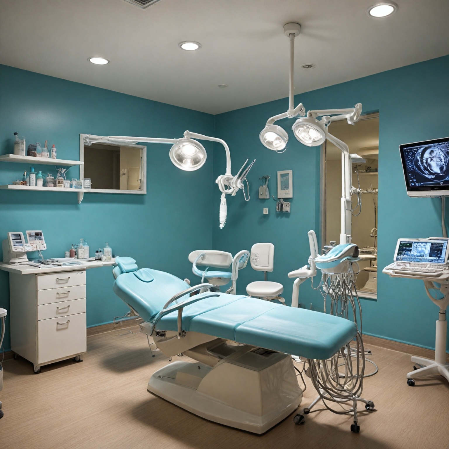
A typical dental implant consists of three main components: the implant post (or screw), the abutment, and the final implant restoration, such as a crown, bridge, or denture. The implant post is surgically inserted into the jawbone and serves as a replacement for the natural tooth root. It is commonly made from biocompatible materials like titanium or zirconium oxide, which comply with international standards set by organizations such as the International Organization for Standardization (ISO) and ASTM International. The implant post usually measures around 18 mm in length and has a tapered end, with a variety of diameters available to accommodate different anatomical requirements, such as replacing smaller teeth like lower incisors. The abutment is a small connector piece that attaches to the top of the implant post and supports the artificial tooth or teeth. Its design varies depending on the implant system used and the type of final restoration intended. For instance, the abutments used to support a single crown differ from those designed to support bridges or dentures. After the implant post has successfully integrated with the bone—a process known as osseointegration—the abutment is placed to enable attachment of the restoration. The implant restoration is the visible portion of the dental implant, typically a crown that is color-matched and shaped to blend with the patient’s natural teeth. In cases where multiple teeth are replaced, bridges or dentures are employed instead of individual crowns. These components work in unison to provide a strong, functional, and aesthetic replacement for missing teeth, with the implant post providing foundational stability, the abutment serving as a connector, and the restoration completing the prosthetic appearance and function.
Materials
Dental implants are fabricated from a variety of materials, each selected based on properties such as durability, biocompatibility, corrosion resistance, and aesthetics. Titanium and its alloys remain the most commonly used materials due to their excellent physical and mechanical strength, chemical stability, high biocompatibility, and ability to integrate with bone through a process known as osseointegration, which is critical for the long-term success of implants. These titanium implants typically consist of three components: the post, abutment, and crown, offering a strong and durable foundation. Ceramic-based materials, including zirconia, zirconia toughened alumina, and alumina toughened zirconia, have gained popularity as alternative biomaterials for dental implants, especially for patients with metal allergies or those seeking improved aesthetics. Zirconia implants are usually one-piece designs, which can simplify the surgical procedure. While polymers have also been explored for dental implant use, they generally lack the durability provided by metals and ceramics. Other materials, such as gold alloys, cobalt-based alloys, titanium alloys, and various ceramics, have also been utilized in dental implant systems. The safety profiles of these materials are well-documented, and they undergo rigorous biocompatibility testing to ensure that bodily contact with the device does not lead to irritation, allergic reactions, or other adverse effects. Surface treatments and coatings, including machined, etched, and sand-blasted surfaces, as well as coatings like hydroxyapatite and fluoride, have been developed to enhance osseointegration and implant longevity. Emerging research also indicates that local administration of agents such as melatonin can improve bone density around implants, further supporting successful osseointegration. Material selection for dental implants takes into account multiple factors including durability, biocompatibility, aesthetics, cost-effectiveness, and patient preferences, all of which contribute to the optimal performance and acceptance of the implant.
Design and Surface Characteristics
The design and surface characteristics of dental implants are critical factors influencing implant stability and the process of osseointegration. Implant geometry, including its macroscopic shape and microscopic surface topography, governs how mechanical stress is distributed to the surrounding bone and plays a significant role in achieving primary and long-term stability.
Impact on Osseointegration and Stability
The process of osseointegration involves several stages: initial incorporation of the implant with woven bone formation, adaptation of bone mass with lamellar and parallel-fibered bone, and long-term bone remodeling in response to functional loading. The surface roughness influences each of these stages by modulating cell adhesion, proliferation, and mineralization. Studies evaluating implant stability using resonance frequency analysis (RFA) and insertion torque (IT) measurements indicate that roughened implant surfaces generally yield better stability outcomes within the first three months after placement compared to smoother surfaces. However, despite evidence supporting the biomechanical benefits of surface roughness, the exact impact of implant microstructure on stability remains inconclusive and is subject to ongoing investigation.
Implant Geometry
The shape of dental implants can be broadly categorized into subperiosteal, transosteal, and endosseous types. Among these, endosseous implants are the most commonly used, and their macroscopic design positively correlates with load distribution and biomechanical stress on the bone site. Implant geometry affects mechanical locking by providing initial stability through features such as ridges, crests, and thread edges, which promote bone growth preferentially on these protrusions. Additionally, the overall shape governs the surface area available for stress transfer and stability, making it a key determinant of implant success.
Surface Topography and Roughness
Surface roughness at both micro- and nanoscales plays a vital role in osseointegration and implant stability. Microroughness, characterized by surface irregularities typically in the micron range, facilitates better adhesion and proliferation of bone cells, directly contributing to improved osseointegration and long-term implant success. Bone embedded in rough-surfaced implants tends to be harder and stiffer compared to bone in contact with machined (smooth) surfaces, indicating enhanced biomechanical quality. Nanoroughness, involving irregularities less than 100 nanometers, also profoundly influences osseointegration by affecting cellular responses at the implant interface. This fine-scale roughness enhances the adsorption of key ions such as calcium (Ca²⁺) and phosphate (PO₄³⁻), which are essential for biomineralization and the subsequent biological integration process.
Biological Process
Osseointegration is the fundamental biological process underlying the success of dental implants, defined as the direct structural and functional connection between living bone and the implant surface. This process ensures the stable anchorage of the implant within the jawbone, providing a foundation for prosthetic restoration and long-term implant stability. The osseointegration process can be divided into three sequential stages. Initially, woven bone forms around the implant, incorporating it into the bone matrix. This is followed by bone mass adaptation, where lamellar and parallel-fibered bone develop in response to mechanical loading. Finally, bone remodeling occurs, refining the bone structure to maintain functional integrity under ongoing biomechanical stress. The entire process resembles fracture healing, wherein immature bone is gradually replaced by mature lamellar bone. The implant surface plays a critical role in osseointegration by influencing the adsorption of calcium (Ca²⁺) and phosphate (PO₄³⁻) ions, which are essential for biomineralization and bone formation. Surface characteristics such as topography and chemistry also modulate the behavior of immune cells, which in turn affects bone regeneration and implant integration. Immunomodulation is therefore a key factor in determining the success of osseointegration, as it governs the local biological environment around the implant. Mechanical factors significantly impact the biological process. Micromotion at the bone-implant interface can damage bone tissue and stimulate cellular signaling pathways, potentially leading to the formation of fibrous tissue rather than direct bone contact. Such fibrous encapsulation can result in implant failure due to compromised osseointegration. Thus, achieving primary stability during implant placement and minimizing micromotion are critical considerations. The concept of osseointegration was initially introduced by Branemark, who described it as a direct connection between living bone and a load-carrying implant at the microscopic level. Subsequent clinical studies, including long-term microscopic observations of human microcirculation in titanium chambers, demonstrated the biocompatibility and absence of inflammatory responses associated with titanium implants. These findings reinforced the feasibility of osseointegration in humans and laid the foundation for modern dental implantology. In some cases, preparatory surgical procedures such as bone distraction may be necessary to enhance the biological environment for implant placement. Bone distraction stimulates new bone growth by gradually separating existing bone segments, allowing sufficient bone volume to support the implant. This process typically requires several months to complete before implantation can proceed.
Surgical Procedure
Dental implant surgery involves several established protocols, primarily categorized into three approaches: two-stage, one-stage, and immediate-loading techniques. The two-stage approach is the most traditional method, where the implant body is first placed below the soft tissue to allow bone healing, typically lasting 2 to 3 months for the mandible and 3 to 6 months for the maxilla. This period facilitates osseointegration—the direct structural and functional connection between living bone and the implant surface, which is critical for implant stability and long-term success. Presurgical planning and communication among the restorative dentist, periodontist, dental technician, and implant surgeon are crucial. This multidisciplinary collaboration ensures that the functional and aesthetic outcomes of the implant-supported rehabilitation are predictable and controlled before the surgery begins. Before the procedure, patients may receive antibiotics and sedative options such as intravenous sedation or nitrous oxide (laughing gas) for comfort. Local anesthesia is administered to numb the surgical site. Depending on the clinical scenario, implants may be restored immediately after placement, although the surgeon typically
Patient Selection
Patient selection is a critical factor in the success of dental implant therapy and involves a comprehensive evaluation of multiple criteria. These include the patient’s age, local anatomical factors, systemic health conditions, educational level, economic considerations, and importantly, the patient’s willingness and compliance to undergo surgery. Thorough history taking and clinical examination are essential to assess these factors and ensure the patient is an appropriate candidate for implantation. Strict patient and site selection criteria have been shown to correlate with high survival rates of dental implants, particularly Type 1A implants, which demonstrate predictable clinical outcomes in partially edentulous patients. However, ongoing research is needed to fully assess esthetic and functional success in these cases. Several relative contraindications must be considered, including cognitive decline, severe systemic conditions classified as American Society of Anesthesiology (ASA) status IV or higher, and other medical issues that may jeopardize patient safety or implant survival. Behavioral and local risk factors such as a history of periodontal disease, bruxism, smoking, and prior radiation therapy also increase the risk of implant failure. Additionally, anatomical considerations—such as the proximity to the mandibular canal, maxillary sinus, nasal floor, and nasopalatine canal—play a significant role in implant planning and patient selection to avoid complications like sinus perforation or nerve injury. Despite the high overall success rate of dental implants, which can reach up to 99%, contraindications related to abnormal anatomical structures or pathological conditions can elevate the risk of failure. Consequently, patient selection not only involves medical and anatomical assessment but also requires careful planning and diagnostic procedures to prevent intraoperative complications and optimize outcomes. Another important aspect is maintaining the integrity of the biologic seal at the permucosal passage—the interface between the implant prosthesis and bone. This area is vulnerable to tissue breakdown that can lead to peri-implantitis and eventual implant failure, emphasizing the need for careful patient and site evaluation to ensure long-term implant longevity.
Clinical Considerations and Challenges
Dental implants require careful clinical consideration to ensure successful outcomes and long-term durability. One of the key factors influencing implant success is the design of the implant itself. Implant shape—generally categorized into subperiosteal, transosteal, and endosseous types—plays a critical role in load distribution and biomechanical stress on the surrounding bone, which in turn affects initial implant stability and osseointegration. Additionally, implant surface characteristics, material selection, and prosthetic design significantly influence both mechanical performance and biological response. Anatomical considerations are paramount during implant planning. Precise evaluation of local structures such as the mandibular canal, maxillary sinus, cortical bone width, and bone density is essential to avoid complications like nerve injury or sinus perforation. For example, to prevent iatrogenic sinus perforation, clinicians may opt for short implants or perform sinus lift and bone augmentation procedures. The choice of surgical protocol—two-stage, one-stage, or immediate-loading—is influenced by these anatomical factors as well as patient-specific conditions, with healing times varying accordingly. Soft tissue management around implants is another critical challenge. The soft tissue acts as a biological “gatekeeper,” preventing bacterial ingress and food impaction that can compromise the implant’s osseous support. Maintaining high-quality peri-implant soft tissue is essential to prevent chronic infections and peri-implant diseases. The permucosal zone between the prosthetic components and bone is particularly vulnerable, as it is the initial site of tissue breakdown leading to potential necrosis and implant failure. Peri-implant mucositis and peri-implantitis remain significant clinical concerns due to their multifactorial etiology, primarily driven by bacterial biofilm accumulation. Risk factors include poor oral hygiene, smoking, uncontrolled diabetes, and local factors such as implant surface and prosthesis design. Diagnosis relies on clinical signs such as inflammation, redness, swelling, and bleeding on probing without radiographic bone loss. Nonsurgical management strategies, including mechanical debridement and photodynamic therapy, are effective in early stages, while surgical intervention may be necessary for more advanced disease. Regular professional maintenance and patient home care are indispensable to minimize these risks and ensure implant longevity. Finally, the clinical phase involving abutment placement and prosthetic restoration is crucial to restore function and aesthetics. The abutment connects the implant fixture to the crown or denture, requiring precise placement after osseointegration to ensure stability and proper load transfer. The entire treatment process, from implant placement to final prosthesis installation, can span from 3 to 6 months, followed by a lifelong maintenance phase that demands continuous monitoring and care. Advancements in materials, implant design, surgical techniques, and digital technologies continue to address existing limitations, aiming to improve clinical outcomes and implant survival rates. However, a comprehensive understanding of patient-specific anatomical, biological, and mechanical factors remains essential for optimal treatment planning and management.
Peri-Implant Diseases
Peri-implant diseases are inflammatory conditions affecting the tissues surrounding dental implants. They are broadly classified into two main entities: peri-implant mucositis and peri-implantitis.
Peri-Implant Mucositis
Peri-implant mucositis is characterized by inflammation confined to the mucosa surrounding an implant without any accompanying loss of supporting bone. Clinical signs typically include bleeding on probing (BoP), redness, and swelling of the mucosa. Diagnosis relies on detecting these signs during regular peri-implant probing at recall visits, without radiographic evidence of bone loss. The primary etiology is bacterial biofilm accumulation, with contributing risk factors such as poor oral hygiene, smoking, uncontrolled diabetes, and implant-specific factors like surface characteristics and prosthesis design. Treatment generally involves nonsurgical approaches focused on mechanical debridement of the implant surface, employing instruments such as curettes, ultrasonic devices, air-abrasive tools, or lasers. Adjunctive use of local antibiotics or antiseptics may also be applied.
Peri-Implantitis
Peri-implantitis represents the progression of peri-implant mucositis to include inflammation of the mucosa and supporting bone, resulting in loss of osseointegration. It is characterized clinically by bleeding and/or suppuration on probing, increased probing pocket depths, mucosal recession, and radiographic evidence of bone loss compared to prior examinations. The condition reflects a breakdown of the biological seal between the implant and the bone, with progressive marginal bone loss compromising implant stability. Established peri-implantitis is challenging to treat predictably, and management often involves a combination of nonsurgical and surgical interventions, though prevention remains the most effective strategy. Prevention emphasizes meticulous personal and professional plaque control to minimize bacterial colonization and inflammation.
Classification and Diagnosis
A classification system based on peri-implant clinical and radiographic parameters stages peri-implant mucositis and peri-implantitis severity. Stage 0A and 0B correspond to peri-implant mucositis, while stages I to IV reflect increasing severity of peri-implantitis. The accepted criteria for implant success, established in 1986, consider annual bone loss of up to 0.2 mm after the first year as normal remodeling, with success rates of 85% at 5 years and 80% at 10 years. Bone loss beyond these thresholds often indicates disease progression.
Prevention and Treatment of Peri-Implant Diseases
Peri-implant diseases are inflammatory conditions affecting the tissues surrounding dental implants and include peri-implant mucositis and peri-implantitis. Peri-implant mucositis is limited to inflammation of the mucosa around the implant, while peri-implantitis involves inflammation that extends to the supporting bone, resulting in loss of osseointegration and increased probing depths, often accompanied by bleeding or suppuration on probing.
Prevention
The primary strategy for managing peri-implant diseases is prevention, which emphasizes maintaining good oral hygiene to minimize bacterial plaque accumulation. Regular personal oral hygiene practices combined with professional cleaning at least twice a year are essential to prevent the onset of peri-implant mucositis and peri-implantitis. Proper brushing technique using an appropriate toothbrush, such as a powered rotary toothbrush, and avoidance of harmful habits like tobacco use and teeth grinding are critical to maintaining implant health. Postoperative care following implant placement or related surgical procedures also plays a vital role in preventing infection and promoting healing, where gentle but thorough cleaning of teeth, including those adjacent to the surgical site, is recommended.
Treatment
Nonsurgical Therapy
Early-stage peri-implant mucositis and mild peri-implantitis cases are often managed with nonsurgical approaches. Mechanical debridement of the implant surface using instruments such as curettes, ultrasonic devices, air-abrasive systems, or lasers is a common practice. These interventions may be supplemented with local application of antibiotics or antiseptics to control infection. Nonsurgical therapy aims to disrupt biofilm formation and reduce inflammation, and in some cases, this approach alone may be sufficient to arrest disease progression.
Surgical Therapy
In cases where peri-implantitis has led to deeper circumferential or intrabony defects, surgical intervention is usually required. The objectives of surgical treatment include thorough debridement, decontamination of the implant surface, and reconstruction of the bone defect when possible. When defects lack well-defined bony walls or predominantly involve suprabony components, surgery focuses on meticulous cleaning and repositioning of the marginal mucosa to facilitate effective oral hygiene by the patient, although this may compromise the esthetic outcome of the implant restoration. The choice of surgical technique depends on the severity and morphology of the defect, and the overall goal is to halt disease progression and maintain implant function.
Postoperative Care
Postoperative care is a critical component in ensuring the success of dental implants following the surgical placement of the implant. Proper adherence to care instructions and attending scheduled follow-up appointments are essential to facilitate healing and to achieve a favorable outcome, including the eventual placement of the abutment and crown that complete the restoration process. Since dental implant placement is a surgical procedure, postoperative care focuses on minimizing pain, swelling, and the risk of infection. Patients are advised to carefully follow instructions to avoid complications, including maintaining good oral hygiene to promote faster healing and reduce discomfort. Neglecting oral hygiene, such as refraining from brushing or rinsing near the surgical site, increases the risk of postoperative wound infection and exacerbated pain. Twenty-four hours after tooth extraction or implant surgery, all teeth—including those adjacent to the surgical site—should be gently brushed without scrubbing to clean areas where food debris may accumulate. Before surgery, patients may receive antibiotics to prevent infection, as well as options for intravenous sedation or nitrous oxide (laughing gas) to increase comfort during the procedure. A local anesthetic is administered to numb the implant site. Depending on the clinical situation, some implants may be restored immediately after placement, while others require a healing period before the abutment or healing cap is placed during a brief follow-up visit. There are three primary surgical protocols for implant placement: two-stage, one-stage, and immediate-loading procedures. The two-stage approach involves placing the implant body below the soft tissue and allowing the bone to heal over a period of typically 2 to 3 months for the mandible and 3 to 6 months for the maxilla before uncovering the implant. Postoperative maintenance also includes preventing peri-implant diseases such as peri-implant mucositis, which can be managed effectively with good oral hygiene and professional dental cleanings at least twice a year. In cases where anatomical defects or compromised tissues are present, surgical interventions may aim to reposition the marginal mucosa to enhance the patient’s ability to maintain hygiene, although this can sometimes affect the esthetic outcome. Emerging adjunctive therapies, such as probiotics, paraprobiotics, and postbiotics, have shown potential in improving clinical outcomes for peri-implant conditions, although further evidence is needed to confirm their effectiveness. Patients are encouraged to avoid poor oral habits such as tobacco use and teeth grinding, which can contribute to implant and prosthetic failure. Utilizing appropriate oral hygiene tools, such as rotary toothbrushes, is recommended to maintain healthy peri-implant tissues and ensure long-term success of the implant-supported restoration.
Risks and Complications
Complications related to dental implants can be broadly classified into accidents occurring during surgery and pathological conditions arising postoperatively. Accidents are intraoperative events that may adversely affect the surgical outcome, while complications are postoperative conditions that can arise early during the healing phase or later during osseointegration. Early-stage complications interfere with the initial healing process, whereas late-stage complications often develop during the implant’s integration with the bone. Local complications during implant surgery play a crucial role in determining the success of the rehabilitation program. Prevention through careful clinical and radiographic examination, accurate surgical planning, the use of appropriate instruments and techniques, and proper management of healing and osseointegration is essential to minimize these risks. Despite a high overall success rate of dental implants (approximately 99%), certain contraindications such as abnormal anatomical structures or pathological conditions can increase the risk of implant failure. Postoperative swelling is a common occurrence following oral surgery, typically peaking within 48 hours. It can be managed by keeping the head elevated and applying cold compresses intermittently during the first two days after surgery. Peri-implant diseases, including peri-implant mucositis and peri-implantitis, represent significant biological complications. Clinical signs of peri-implantitis include inflammation evidenced by bleeding upon probing, suppuration, increased probing pocket depths, mucosal recession, and radiographic bone loss compared to earlier examinations. Management strategies aim primarily at eliminating bacterial contamination and detoxifying the implant surface. Treatment modalities range from nonsurgical mechanical debridement using curettes, ultrasonic devices, air-abrasive devices, or lasers to adjunctive use of local antibiotics or antiseptics, and may extend to surgical intervention depending on disease severity. Systemic antibiotics combined with debridement have also been studied for managing peri-implant mucositis, with evidence supporting their use in certain cases. Technical complications, often termed “prosthetic complications,” affect both fixed and removable implant-supported prostheses. These issues include mechanical failures and material-related problems linked to implant design, surgical technique, bone quality and quantity, occlusal forces, and implant surface characteristics. Prevention and management of these complications require thorough understanding of implant biomechanics and meticulous prosthetic planning.
Advantages and Benefits
Dental implants offer several significant advantages over traditional tooth replacement options. One of the primary benefits is the improvement in oral function, allowing patients to eat, speak, and smile with greater confidence and comfort. This enhanced functionality is largely due to the stable foundation implants provide, as the implant surface fuses with the surrounding bone through a process known as osseointegration, ensuring long-term stability within the oral environment. The popularity and increasing use of dental implants are influenced by multiple factors, including psychological impacts of tooth loss, an aging population, and the limitations of removable prostheses. Implants provide predictable long-term results that improve quality of life by restoring both function and aesthetics more effectively than other prosthetic solutions. Additionally, considerations such as durability, biocompatibility, aesthetics, and cost-effectiveness play crucial roles in selecting implant materials that meet individual patient needs and preferences. Moreover, advancements in implant materials, design modifications, surgical techniques, and digital technologies continue to enhance the efficacy and longevity of dental implants, addressing previous limitations and further improving patient outcomes. These improvements, combined with personalized care and modern surgical resources, contribute to high-quality treatment results and an overall positive patient experience. Surgical interventions associated with implant placement and maintenance also aim to ensure thorough debridement and implant surface decontamination, particularly in cases involving peri-implant defects. Such interventions help maintain the health and functionality of the implant, although in some cases, measures to enable effective oral hygiene may slightly compromise esthetic outcomes.
Cost and Insurance Considerations
The cost of dental implants can vary significantly depending on several factors, including the type of implant used—such as endosteal, subperiosteal, or zygomatic—and the material, typically titanium or other alternatives. Geographic location and the complexity of the procedure also influence the overall price. For example, dental implant costs in Scottsdale are subject to these variables, making it essential for patients to receive a tailored cost estimate from their dental provider. Insurance coverage for dental implants is inconsistent and often limited. Many insurance plans do not fully cover implant procedures, and coverage may vary widely between providers and policies. Patients are advised to verify their specific insurance benefits before proceeding to understand potential out-of-pocket expenses. Unlike other dental restorations, implants may require more frequent maintenance and occasional replacements, which could incur additional costs over time. To maximize the success and longevity of implants—and potentially reduce further expenses—patients are encouraged to follow thorough aftercare instructions, such as consuming soft foods, maintaining good oral hygiene, and avoiding smoking. Regular follow-up visits allow dental professionals to monitor healing and address any complications promptly. Preventing surgical complications through careful clinical and radiographic examination, precise planning, and the use of appropriate surgical techniques also contributes to reducing unforeseen costs and improving rehabilitation outcomes.
Alternatives to Dental Implants
While dental implants offer a durable and natural-looking solution for tooth loss, there are several alternatives available for patients who may not be suitable candidates for implants or prefer different options. These alternatives primarily include dentures and dental bridges. Dentures are removable prosthetic devices designed to replace missing teeth and surrounding tissues. They can be full, replacing all teeth in an arch, or partial, filling in gaps where some natural teeth remain. Dentures are often a more affordable option compared to implants and do not require surgery. However, they may be less stable and require regular maintenance. Dental bridges involve the placement of crowns on adjacent teeth to support a false tooth or teeth in between. Unlike implants, bridges rely on neighboring natural teeth for support rather than anchoring directly into the jawbone. Bridges can provide a fixed solution without surgery, but they may require the alteration of healthy teeth and do not prevent bone loss in the area of the missing tooth. Other less common alternatives include resin-bonded bridges and removable partial dentures, which offer different levels of permanence and invasiveness depending on the patient’s needs. Choosing the best alternative depends on various factors such as oral health, bone density, budget, and personal preferences. Consulting with a dental professional is essential to determine the most suitable treatment plan.
Research and Future Developments
Recent advancements in dental implantology focus on overcoming existing limitations while improving the efficacy, longevity, and biocompatibility of implants. Innovations in implant surface treatments such as acid etching, sandblasting, and plasma spraying have significantly enhanced osseointegration, making implants more reliable and durable. Surface coatings like hydroxyapatite, fluoride, and biodegradable compounds containing silicon have been developed to promote bone generation and improve integration. Newer coatings such as Laminin I offer the potential to enhance osseointegration while maintaining smooth implant surfaces, thereby reducing complications related to rough textures. Materials science continues to play a crucial role in implant development. Titanium and its alloys remain the gold standard due to their favorable biocompatibility and physical properties, but ceramic-based materials—including zirconia and alumina composites—are gaining traction as promising alternatives that may offer improved aesthetics and reduced allergic reactions. Furthermore, the immunomodulatory effects of implant surface topography have been recognized as critical factors influencing bone healing and osseointegration, highlighting the interplay between implant design and the host’s biological response. Technological advancements are also transforming surgical techniques and implant design. Digital technologies have revolutionized planning and placement procedures, allowing for greater precision and customized solutions across dental specialties such as prosthodontics, periodontics, orthodontics, and pediatric dentistry. Notably, pediatric implants demonstrate a high success rate of 90-95% over ten years, emphasizing the potential for broader applications. Emerging fields like 4D printing promise dynamic, adaptable implants capable of responding to physiological changes over time, representing a frontier in personalized dental care. On the biological front, adjunctive therapies are being explored to enhance osseointegration. For example, local melatonin administration has been shown to increase bone density around immediately loaded implants, suggesting new avenues for improving implant stability and success rates. Continued research into immune cell interactions with implant surfaces aims to refine implant designs that actively promote favorable healing responses while minimizing inflammation.
“`
The content is provided by Blake Sterling, Gear Shift Zone