Highlights
- Severe spinal stenosis at L4-L5 significantly impairs mobility and quality of life.
- Minimally invasive surgery offers substantial pain relief and faster recovery for affected patients.
Summary
Severe spinal stenosis at the L4-L5 level is a debilitating condition characterized by the narrowing of the spinal canal in the lower lumbar region, leading to compression of spinal nerves and significant neurological symptoms. This level is the most frequently affected segment due to its high mechanical stress and susceptibility to degenerative changes such as disc herniation, bone spurs, and ligament thickening. Patients often present with chronic lower back pain, leg numbness, weakness, and in severe cases, complications like foot drop and cauda equina syndrome, which can severely impair mobility and quality of life.
Lumbar spinal stenosis (LSS) at L4-L5 is notably prevalent among older adults and is one of the leading causes of spine-related disability and surgical intervention worldwide. Diagnosis relies on a combination of clinical assessment and imaging modalities—primarily MRI—which reveal critical canal narrowing and nerve root compression. Conservative treatments, including physical therapy, medication, and epidural steroid injections, form the initial management strategy and can effectively alleviate symptoms for many patients. However, surgery becomes necessary when conservative measures fail or neurological deficits progress.
Surgical options have evolved significantly, with minimally invasive techniques increasingly preferred to reduce recovery time and complications while effectively decompressing the spinal canal. Despite surgical risks, timely intervention often results in substantial improvement in pain and function, particularly when patients are carefully selected and managed by multidisciplinary spine care teams. Ongoing research continues to refine both conservative and surgical approaches, emphasizing personalized treatment plans that balance efficacy and safety.
Controversies remain regarding optimal management, particularly the comparative long-term benefits of conservative versus surgical therapies and the role of advanced minimally invasive procedures. Additionally, variability in patient response to exercise-based interventions and the complexity of surgical decision-making underscore the need for further high-quality research to guide individualized care.
Anatomy and Physiology of the Lumbar Spine
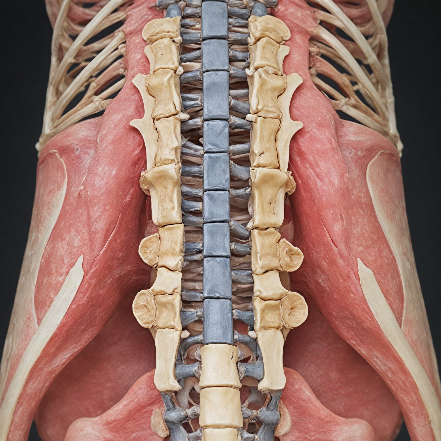
The lumbar spine consists of five vertebrae labeled L1 through L5, forming the lower part of the spine that supports much of the body’s weight and allows for a wide range of motion. Among these, the L4-L5 segment is the most commonly affected level in lumbar spinal stenosis (LSS), followed by L3-L4, L5-S1, and L1-L2. This region endures significant mechanical stress during daily activities, making it particularly vulnerable to degenerative changes such as disc herniation, bone spurs, and ligament thickening. The spinal canal within the lumbar spine houses the spinal cord and nerve roots. In LSS, this canal narrows, often due to degenerative processes like bulging discs or thickened ligaments, which can compress spinal nerves and lead to symptoms. The spinal nerve roots at the L4-L5 level are critical as they supply sensation and motor function to various parts of the lower body. Compression at this level can impinge on the L5 nerve root, causing symptoms such as sciatica—a sharp, radiating pain along the leg. Furthermore, the cauda equina, a bundle of nerve roots located between L1 and L5, may also be affected by nerve compression at L4-L5, increasing the risk of cauda equina syndrome, a serious neurological condition.
Overview of Spinal Stenosis
Spinal stenosis is a medical condition characterized by the narrowing of the spinal canal, which results in the compression or pinching of the spinal nerves or spinal cord. This narrowing can occur anywhere along the spine but most commonly affects the neck (cervical spine) and the lower back (lumbar spine). In particular, lumbar spinal stenosis (LSS) at the L4-L5 level is a frequent site of degeneration due to the significant mechanical stress this region endures during daily activities.
The pathophysiology of spinal stenosis typically involves degenerative changes such as hypertrophic lesions of the facet joints, thickening of the ligamentum flavum, bone spur (osteophyte) formation, and intervertebral disc bulging or herniation. These changes lead to a reduction in the space available for nerve roots and blood vessels, causing debilitating compression symptoms. Spinal degeneration related to aging is the most common cause, with osteoarthritis contributing to facet joint changes and disc degeneration, thereby increasing the risk of stenosis.
Clinically, patients with spinal stenosis often experience symptoms including persistent lower back pain, numbness, tingling, and muscle weakness in the legs or arms, depending on the site of compression. In severe cases, symptoms may escalate to significant neurological deficits such as difficulty walking or problems with bladder and bowel control, underscoring the importance of early recognition and intervention. Radiologically, spinal stenosis is identified by a narrowed spinal canal diameter, with criteria such as midsagittal diameters less than 12 mm indicating relative stenosis and less than 10 mm representing absolute stenosis.
Spinal stenosis is notably prevalent among the elderly, with radiological studies indicating that moderate to severe stenosis can affect up to 80% and 40% of individuals over 40 years of age, respectively. Genetic factors have also been implicated in the pathogenesis, influencing osteophyte formation and ligamentous hypertrophy. Due to its high prevalence and disabling nature, spinal stenosis remains one of the most common indications for spinal surgery in patients older than 65 years.
Clinical Presentation
Severe spinal stenosis at the L4-L5 level manifests through a variety of symptoms that significantly impact a patient’s daily life and mobility. The hallmark signs include chronic lower back pain, radiating discomfort into the legs, tingling, numbness, and muscle weakness, which often intensify during activities such as walking or standing but may improve with bending forward or sitting. Patients frequently report difficulty walking more than short distances without needing rest, as well as challenges maintaining balance or standing upright for prolonged periods.
A common neurological complication associated with L4-L5 stenosis is foot drop, characterized by an inability to properly lift the front part of the foot during walking, causing the foot to slap onto the ground and increasing the risk of falls. This condition results from nerve root compression and can severely impair mobility; however, with prompt treatment including physical therapy, bracing, and nerve stimulation, many cases improve and are not permanent.
In more severe cases, nerve involvement may progress to cause significant muscle weakness, loss of sensation, and impaired reflexes. These neurological deficits can escalate to cauda equina syndrome, a medical emergency marked by bladder or bowel dysfunction that necessitates immediate intervention to prevent permanent damage and loss of independence.
Diagnosis typically involves a thorough physical examination to detect symptoms such as numbness, weakness, and altered reflexes in various positions, followed by imaging studies like MRI or CT scans to assess the degree and exact location of spinal canal narrowing. Additional tests such as electromyography (EMG) may be used to evaluate nerve function. Early recognition of these clinical signs is crucial for timely management and prevention of irreversible nerve damage.
Diagnostic Evaluation
The diagnostic evaluation of severe spinal stenosis at the L4-L5 level involves a comprehensive clinical and imaging assessment to accurately identify the presence and severity of stenosis, as well as its impact on neurological function. Initially, a detailed medical history and symptom review are essential, focusing on characteristic complaints such as leg numbness, weakness, atypical reflexes, and frequent calf cramping, which often worsen with walking or at night due to nerve root compression. A thorough physical examination assesses these neurological signs and helps differentiate spinal stenosis from other causes of low back pain.
Imaging studies play a pivotal role in confirming the diagnosis and guiding treatment decisions. Magnetic resonance imaging (MRI) of the lumbosacral spine is considered the modality of choice due to its superior soft tissue resolution, enabling sensitive detection of spinal nerve lesions and detailed visualization of the degree of canal narrowing. Typical MRI findings for L4-L5 stenosis include narrowing of the central spinal fluid signal on T2-weighted images and evidence of central canal stenosis. Computed tomography (CT) scans may complement MRI by providing more detailed images of bony structures and osteophyte formation that contribute to stenosis. Radiographic criteria often used to define severity include an intraspinal canal area less than 76 mm² for severe stenosis and anteroposterior spinal canal diameters under 10 mm. The nerve root sedimentation sign on MRI further aids in diagnosis, with its absence indicating spinal stenosis.
Additional diagnostic tools such as electromyography (EMG) may be utilized to assess nerve function and differentiate radiculopathy from other neuropathies. Plain X-rays, although less sensitive, provide valuable information about spinal alignment, intervertebral disc height, and potential instability, which are important considerations before surgical intervention. Dynamic imaging techniques may also be employed to evaluate spinal stability and the necessity for decompression or fusion procedures.
Importantly, the diagnosis of severe spinal stenosis is not solely based on imaging findings but also integrates the patient’s symptom severity and functional impairment, including neurological deficits like muscle weakness or loss of bowel and bladder control. This comprehensive evaluation is crucial to differentiate patients who may benefit from conservative management, such as physical therapy, from those requiring timely surgical intervention.
Management Strategies
Management of severe spinal stenosis at the L4-L5 level involves a multifaceted approach tailored to the individual patient’s symptoms, severity of the condition, and overall health status. Treatment typically begins with conservative measures aimed at symptom relief and functional improvement, progressing to surgical options when necessary.
Conservative Treatment
Early management emphasizes non-operative interventions, including physical therapy, medications, lifestyle modifications, and injections. Physical therapy plays a central role by focusing on core strengthening, lumbar flexion exercises, and improving spinal flexibility to alleviate nerve compression and enhance spinal support. Core strengthening helps improve posture and spinal alignment, which may reduce symptoms and improve daily function. Aerobic exercises are often incorporated to increase tolerance for activities affected by stenosis, such as walking.
Medications, while primarily used to manage pain and enable participation in physical therapy, have limited long-term effectiveness and carry potential side effects. Epidural steroid injections may be employed to reduce inflammation and provide temporary symptom relief. Conservative treatment is generally pursued for at least a year or until symptoms significantly impair quality of life or neurological function.
Several studies have demonstrated that physical therapy can be as effective as surgery in improving pain and function for spinal stenosis patients, with fewer risks and complications. For example, one study published in the Annals of Internal Medicine found comparable long-term outcomes between patients treated with physical therapy and those undergoing surgery, with the added benefit of fewer surgical complications in the physical therapy group.
Surgical Intervention
Surgery is considered when conservative treatments fail to provide sufficient symptom relief or when patients develop progressive neurological deficits such as significant leg weakness, difficulty walking, or loss of bladder or bowel control. Preoperative evaluation includes a thorough history, physical examination, and imaging studies such as MRI or CT scans to assess the degree of spinal canal narrowing and identify structural contributors like bone spurs.
Minimally invasive surgical techniques are increasingly favored for their potential to reduce recovery time and complications. For example, a combined approach involving minimally invasive fusion and tumor resection at L5-S1, along with a minimally invasive laminectomy at L4-L5, has been successfully employed in complex cases. Surgical options include bilateral or unilateral laminotomy, partial facetectomy, and split-spinous process laminotomy or laminoplasty, with the choice tailored to the patient’s anatomy and clinical presentation.
While surgery carries risks such as infection and incomplete symptom resolution, these are often outweighed by the benefits in patients with severe, disabling stenosis. Successful outcomes depend on careful patient selection, surgical expertise, and comprehensive perioperative care.
Role of Specialist Care and Rehabilitation
Engagement with spine specialists and multidisciplinary teams is critical for optimal management. Centers specializing in spine health offer expert evaluation, personalized exercise programs, and guidance through conservative or surgical treatments. Postoperative rehabilitation typically includes physical therapy aimed at restoring strength, flexibility, and function while minimizing pain and preventing complications.
Prognosis and Outcomes
Early recognition of symptoms in severe spinal stenosis at L4-L5 is crucial for timely intervention, which can significantly improve quality of life and potentially prevent further nerve damage. Delayed treatment may lead to worsening mobility, loss of independence, and decreased overall well-being. Conservative management, such as medication, physical therapy, and injections, is typically the first approach, aiming to relieve symptoms and halt disease progression.
Physical therapy has been shown to be an effective initial treatment modality. Studies indicate that patients who engage in physical therapy within the first six weeks of symptom onset report higher self-rated improvements at 3 to 6 months and up to one year later. Moreover, a randomized trial demonstrated that physical therapy outcomes are comparable to surgical interventions in terms of pain reduction and functional improvement over a two-year period, with the added advantage of fewer complications.
When conservative treatments fail or neurological symptoms become severe—such as significant weakness, impaired walking ability, or bladder control issues—surgical intervention is considered. Surgical options range from decompression alone to decompression combined with instrumented fusion. Evidence suggests that decompression with fusion may provide superior outcomes in terms of disability and pain relief compared to decompression alone, with a meta-analysis indicating it to be approximately 3.5 times more effective in improving Oswestry Disability Index scores and visual analog pain scales for both back and leg pain. However, patient satisfaction may vary, with some studies showing slightly higher satisfaction rates following decompression alone.
Minimally invasive spine surgery is an option for selected patients who have well-defined pathology and are in good overall health. This approach offers the benefits of reduced surgical risks such as infection and hardware complications while providing effective symptom relief. It is important to weigh these benefits against potential risks in a personalized treatment plan developed by experienced neurosurgical teams to achieve optimal outcomes.
Prevention
Preventing severe spinal stenosis at the L4-L5 level involves early recognition of symptoms and timely intervention to avoid progression and complications. Understanding the severity of symptoms and seeking appropriate care can help maintain mobility, independence, and overall quality of life. While some risk factors, such as age and anatomical predisposition, cannot be modified, proactive management strategies can reduce symptom severity and delay or prevent the need for invasive treatments.
Exercise plays a key role in prevention and management. Although there is no single best exercise for spinal stenosis, gentle stretching routines—such as the knee-to-chest stretch—can relieve pressure on the lower back and improve flexibility. However, exercise interventions are complex, and their effectiveness varies based on individual patient factors and intervention components. Current research emphasizes the need for personalized exercise programs tailored to the patient’s condition, symptoms, and response to treatment.
Maintaining spinal health also involves addressing lifestyle factors that contribute to spinal degeneration, including maintaining a healthy weight, practicing good posture, and avoiding activities that exacerbate back stress. Regular monitoring through imaging techniques, especially non-contrast MRI, enables early detection of spinal changes before severe nerve compression occurs.
For individuals with existing symptoms, engaging with specialized spine care providers can ensure comprehensive evaluation and the implementation of personalized prevention and treatment plans. Early intervention, combining conservative management approaches such as physical therapy, medication, and lifestyle modification, may reduce the progression of stenosis and improve long-term outcomes.
Research and Future Directions
Research on managing severe spinal stenosis at L4-L5 continues to evolve, highlighting the complexity of treatment approaches and the need for individualized patient care. Systematic reviews have underscored challenges in interpreting heterogeneous data from exercise interventions, emphasizing that conclusions drawn may have limited utility for clinicians and policymakers aiming to understand how specific exercise components affect patient outcomes
The content is provided by Jordan Fields, Gear Shift Zone